|
Anatomy |
|
Anatomy
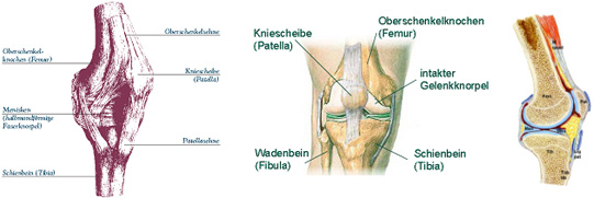
The knee, which is the largest joint of the human body, connects the femur with the tibia and it primarily allows us to stretch and bend our legs.
The healthy knee is incredibly flexible, yet also very stable and durable. Because of it, we can get up, stand and walk.
|
In the knee the femoral condyles and the head of the tibia form the joint. They are covered by cartilage and separated by a junction that holds the menisci.
An additional joint inside the knee is formed by the patella, also called the kneecap, and the femur. The kneecap is the largest sesamoid bone of the human body and it transfers the load of the femoral muscles to the tibia.
A joint capsule encloses the knee joint, and it is filled with a fluid that keeps the cartilage nourished and joint movements smooth. The ligaments together with the muscles provide proper guidance and stability.
The cruciate and lateral ligaments as well as the menisci have sensors that track the state of tension in the tendons. If the tendons are overly stretched, the sensors set in motion a protective reflex that tightens the leg muscles.
|
|
|